Skip Navigation
Skip to contents

ACC : Acute and Critical Care

OPEN ACCESS
SEARCH
Search

Articles

Page Path
HOME > Acute Crit Care > Volume 39(1); 2024 > Article
Original Article
Pediatrics
Outcomes of extracorporeal membrane oxygenation support in pediatric hemato-oncology patients
Hong Yul An1,2orcid, Hyoung Jin Kang1,2orcid, June Dong Park1orcid
Acute and Critical Care 2024;39(1):108-116.
DOI: https://doi.org/10.4266/acc.2023.01088
Published online: January 24, 2024

1Department of Pediatrics, Seoul National University College of Medicine, Seoul, Korea

2Cancer Research Institute, Seoul National University College of Medicine, Seoul, Korea

Corresponding author: June Dong Park Department of Pediatrics, Seoul National University College Of Medicine, 103 Daehak-ro, Jongno-gu, Seoul 03080, Korea Tel: +82-2-2072-3359 Fax: +82-2-743-3455 E-mail: jdparkmd@snu.ac.kr
• Received: August 23, 2023   • Revised: December 13, 2023   • Accepted: December 13, 2023

© 2024 The Korean Society of Critical Care Medicine

This is an Open Access article distributed under the terms of the Creative Commons Attribution Non-Commercial License (http://creativecommons.org/licenses/by-nc/4.0/) which permits unrestricted non-commercial use, distribution, and reproduction in any medium, provided the original work is properly cited.

  • 574 Views
  • 38 Download
  • Background
    In this study, we reviewed the outcomes of pediatric patients with malignancies who underwent hematopoietic stem cell transplantation (HSCT) and extracorporeal membrane oxygenation (ECMO).
  • Methods
    We retrospectively analyzed the records of pediatric hemato-oncology patients treated with chemotherapy or HSCT and who received ECMO in the pediatric intensive care unit (PICU) at Seoul National University Children’s Hospital from January 2012 to December 2020.
  • Results
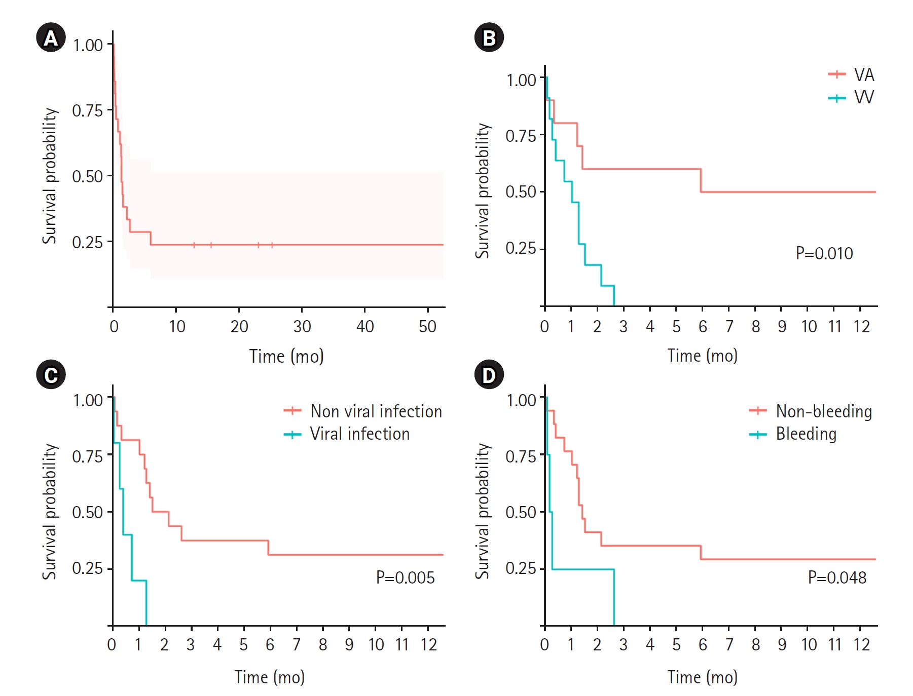
    Over a 9-year period, 21 patients (14 males and 7 females) received ECMO at a single pediatric institute; 10 patients (48%) received veno-arterial (VA) ECMO for septic shock (n=5), acute respiratory distress syndrome (ARDS) (n=3), stress-induced myopathy (n=1), or hepatopulmonary syndrome (n=1); and 11 patients (52%) received veno-venous (VV) ECMO for ARDS due to pneumocystis pneumonia (n=1), air leak (n=3), influenza (n=1), pulmonary hemorrhage (n=1), or unknown etiology (n=5). All patients received chemotherapy; 9 received anthracycline drugs and 14 (67%) underwent HSCT. Thirteen patients (62%) were diagnosed with malignancies and 8 (38%) were diagnosed with non-malignant disease. Among the 21 patients, 6 (29%) survived ECMO in the PICU and 5 (24%) survived to hospital discharge. Among patients treated for septic shock, 3 of 5 patients (60%) who underwent ECMO and 5 of 10 patients (50%) who underwent VA ECMO survived. However, all the patients who underwent VA ECMO or VV ECMO for ARDS died.
  • Conclusions
    ECMO is a feasible treatment option for respiratory or heart failure in pediatric patients receiving chemotherapy or undergoing HSCT.
Extracorporeal membrane oxygenation (ECMO) is a cornerstone intervention for the management of adult and pediatric severe cardiac or respiratory failure that is unresponsive to conventional treatment [1,2]. ECMO is used to supply oxygen, assist with clearance of CO2, and provide circulatory support, creating an opportunity for resuscitation and reducing organ damage from treatment [3]. The use of ECMO in children with certain conditions, especially malignant disease and organ transplantation, poses a high risk of mortality [4]. In pediatric oncology, application of ECMO is challenging due to the unique pathophysiological aspects and complications associated with malignancy and its treatment. The performance of ECMO in children with malignant disease is controversial, and the reported mortality rate ranges from 25%–100%, the latter in a group of children undergoing hematopoietic stem cell transplantation (HSCT) [5,6].
In one multicenter survey, 78% of doctors did not consider malignancy a contraindication for ECMO, 17% considered it a relative contraindication, and 5% an absolute contraindication [7]. Pediatric patients with cancer often present with complications, such as infection, bleeding, and organ dysfunction, which can complicate ECMO management. The type of underlying malignancy, stage of the disease, and associated complications may play significant roles in determining survival outcomes in pediatric patients. In the present study, we aimed to investigate these factors and their effects on ECMO survival outcomes, specifically in pediatric patients with malignancies and subjects undergoing HSCT.
We retrospectively analyzed the medical records of all 21 children treated with chemotherapy or HSCT and who received ECMO in the pediatric intensive care unit (PICU) at Seoul National University Children’s Hospital from January 2012 to December 2020. The Institutional Review Board of Seoul National University waived the need to obtain ethics approval for the study protocol and written consent from the study participants (No. H-2108-122-1246).
Continuous variables are presented as means±standard deviations if normally distributed and as medians (ranges/interquartile ranges [IQRs]) if non-normally distributed. Categorical variables are presented as frequencies (percentages). Statistical analysis was conducted using R software (ver. 3.3.2 GUI 1.68 Mavericks build; The R Foundation for Statistical Computing). The Kaplan-Meier method was performed for analysis of event-free and overall survival, and the log-rank test was used for subgroup comparisons. A P<0.05 was considered statistically significant.
Clinical Characteristics
The mean age of the 21 patients was 4.97 years, 13 patients (62%) had an underlying malignant disease, and 9 patients (43%) previously used doxorubicin. HSCT was performed in 14 patients (67%); 7 (50%) experienced graft-versus-host disease. During treatment, 12 patients (57%) underwent continuous renal replacement therapy and 20 (95%) received ventilation; 5 (25%) received high-frequency ventilation. Five patients (24%) underwent cardiopulmonary resuscitation before EMCO, and ECMO was initiated 75 days on average after anticancer drug administration and 4 days after ventilator application. On average, ECMO was performed for 22 days. Infection was confirmed in 13 patients (62%), some testing positive for 2 or more types of bacteria. Six of 17 patients (35%) were administered 3 or more inotropic agents. The mean oxygen saturation index was 22.17, the mean partial pressure of carbon dioxide (PCO2) was 77.62 mm Hg, mean pH was 7.18, and mean lactic acid concentration was 1.5 mmol/L. The demographic and key clinical characteristics of the 21 patients are summarized in Tables 1 and 2.
Outcomes
Among the 21 patients, two (10%) underwent veno-venous (VV) to veno-arterial (VA) ECMO conversion due to aggravation of cardiac function, one (5%) underwent VA to VV ECMO conversion to improve cardiac function, and two (10%) underwent lung transplantation (Table 3). The mean number of continuous ECMO days was 22.0, ranging from 6.0–42.0 days. Malignant relapse after ECMO occurred in four of 13 patients (31%) with malignant disease. The ICU and hospital survival rates were six of 21 (29%) and five of 21 (24%), respectively. The mean survival was 38.0 days, with a range of 11–180 days. Among the 16 patients who died during their hospital stay, five (31%) died of infection, four (25%) due to bleeding, two (13%) to heart failure, four (25%) of unknown causes, and one (6%) because the family requested ECMO be discontinued. Regarding ECMO complications, infection occurred in seven of 21 cases (33%), bleeding in four of 21 (19%), heart failure in two of 21 (10%), and thromboembolism in four of 21 (19%).
Analyses
Between survivors (n=5) and non-survivors (n=16), significant differences were observed in the number of days ECMO was provided with a ventilator (0.8±1.5 vs. 9.9±9.4, P=0.002), percentage of patients receiving VV ECMO (0% vs. 69%, P=0.030), left ventricular ejection fraction (29% [IQR, 24%–50%] vs. 69% [IQR, 59%–76%], P=0.002), and percentage of patients administered more than three types of inotropics (80,0% vs. 12.5%, P=0.019), and PCO2 (51.4±18.3 mm Hg vs. 85.8±26.4 mm Hg, P=0.014) (Table 4).
Among the 21 participants, 6 had heart-related morbidities (heart group) and 15 had lung-related morbidities (lung group) (Table 5). Comparison of the two groups resulted in several notable observations. The median (IQR) age of participants in the heart group was 9 years (6–16 years, whereas that in the lung group was 4 years (2–6 years, P=0.045). Regarding ECMO modes, all six participants in the heart group underwent VA ECMO, and 73% subjects in the lung group underwent VV ECMO (P=0.011). Regarding oxygenation measures, the mean oxygen saturation was significantly higher in the heart group (87.3%±17.0% vs. 59.5%±21.2%, P=0.010) and the median oxygenation saturation index was significantly higher in the lung group (28.8 [IQR, 19.7–43.1] vs. 6.7 [IQR, 6.2–9.3], P=0.001). The mean hemoglobin concentration was lower in the heart group (9.1±1.8 g/dl) than in the lung group (11.0±1.4 g/dl, P=0.016). In blood gas analyses, the median PCO2 level was significantly higher in the lung group (94.0 mm Hg [IQR, 70.5–114.5 mm Hg] vs. 41.0 mm Hg [IQR, 35.0–62.0 mm Hg], P=0.001). The median lactic acid level was significantly higher in the heart group (11.0 mmol/L [IQR, 6.5–15.0 mmol/L] vs. 1.1 mmol/L [IQR, 0.8–1.7 mmol/L], P=0.004) and the median survival time was significantly longer (432.0 days [IQR, 180.0–702.0 days] vs. 30.0 days [IQR, 8.0–41.5 days], P=0.004). The ICU survival rate was significantly higher in the heart group (83% vs.7%, P=0.003) and similar to the hospital discharge rate (67% vs. 7%, P=0.019).
In our analysis of survival curves (Figure 1), we observed a significant divergence between the groups undergoing VV ECMO and VA ECMO, as evidenced by the log-rank test P-value of 0.010 (Figure 1B). This indicated a statistically significant difference in survival rates between these two patient groups. Furthermore, infection type appeared to influence survival outcomes. Specifically, patients with viral infections exhibited a worse survival trajectory than subjects without viral infections (P=0.005) (Figure 1C). This difference may be because viral infections more frequently manifested as respiratory failure than non-viral infections. We also evaluated the effect of complications on patient survival. Notably, patients with bleeding complications had a significantly worse survival curve than those without bleeding (P=0.048) (Figure 1D, Supplementary Figure 1).
Previous case reports have described ECMO in conjunction with various cancer-related treatments. In one study, ECMO was applied after bone marrow transplantation in patients with severe combined immunodeficiency [8], and in another study, ECMO was used in a patient with post-HSCT diffuse alveolar hemorrhaging [9]. In addition, ECMO has been used to treat tumor lysis syndrome, fungemia, and respiratory syncytial virus infection, all of which occur during the treatment of children with leukemia [10,11].
Advances in medical technology have led to improvements in the application of ECMO in pediatric patients with cancer. Recent reports have indicated an ECMO success rate in pediatric settings of 20%–40% [12,13]. In the present study, we observed ICU and hospital survival rates of 29% and 24%, respectively, similar to previously reported survival rates [14-17]. In a previous analysis conducted at a single institution in South Korea, the survival rate among 15 adult patients with hematological malignancies and receiving ECMO was 0% [18]. An academic committee has recently issued guidelines on the application of ECMO for patients undergoing HSCT [19,20]. These guidelines suggest that ECMO should not be considered a contraindication for adult or children HSCT patients. Instead, a multidisciplinary approach should be adopted. The guidelines emphasize the importance of carefully considering the patient's underlying conditions and the potential for relapse when making decisions.
In the present study, the 1-year survival was 38% (95% confidence interval, 0.21–not available). Among patients treated for septic shock, three of five (60%) who underwent ECMO and five of 10 (50%) who underwent VA ECMO survived. However, all the patients who underwent VA ECMO or VV ECMO for acute respiratory distress syndrome died. ECMO for circulatory failure yielded significantly better results than ECMO for respiratory failure, which might have been due to the significantly older age and rapid application of ECMO from the time of initiation of ventilator care in the latter group. Furthermore, in VA ECMO cases, the pathogen was identified relatively quickly, and appropriate antibiotics were available. However, in VV ECMO cases, most patients were affected by viruses, rendering ineffective the use of antibiotics. Maintaining ECMO was also challenging in several conditions such as bleeding, which could explain the outcomes observed. Based on the results, lower ejection fraction, lower blood pressure, higher lactic acid concentration, and use of three or more types of inotropics may paradoxically contribute to survival, likely because VA ECMO resulted in better survival than VV ECMO. To the best of our knowledge, a direct comparison between the outcomes of VV ECMO and VA ECMO has not been reported; however, a survival rate of 44% was observed among pediatric patients with neutropenic sepsis who underwent VA ECMO [21].
In the present study, two of five patients (40%) undergoing HSCT survived ECMO, which is higher than the 20% survival rate reported in previous studies involving similar patients [22,23]. The most common complications of ECMO in the present study were infection and bleeding, and the incidence of thrombus formation was significantly higher with VA ECMO than with VV ECMO. Notably, 80% of post-infection deaths appeared attributable to imipenem-resistant Acinetobacter baumannii. Because patients with viral infections had worse survival trajectories than those without viral infections, the results underscore the influence of infection type on survival outcomes. Cytomegalovirus infections frequently led to respiratory failure in our study. This observation is consistent with the established understanding of the severe respiratory implications of certain viral infections. Furthermore, the presence of bleeding complications was associated with significantly worse survival rates, consistent with existing literature emphasizing the impact of bleeding complications in critical care settings, especially in patients receiving ECMO. Therefore, efforts should be made to predict, prevent, and promptly manage bleeding complications in high-risk patients. Further investigations are needed to determine the exact factors that predispose patients on ECMO to bleeding, ranging from anticoagulation management to patient-specific characteristics.
In conclusion, our results highlight the potential avenues for future research and opportunities for clinical practice improvements in ECMO management. The type of ECMO strategy used, nature of underlying infections, and management of complications, specifically bleeding, may significantly affect survival outcomes. Such insights are critical for patient-centered, evidence-based critical care in the era of ECMO. However, these results should be interpreted in consideration of the study design and context, and further studies should be performed in diverse settings and with larger patient cohorts.
▪ Six of 21 patients (29%) survived extracorporeal membrane oxygenation (ECMO) in the pediatric intensive care unit and 5 patients (24%) survived to hospital discharge.
▪ ECMO is a feasible treatment option for respiratory or heart failure in pediatric patients receiving chemotherapy or undergoing hematopoietic stem cell transplantation.

CONFLICT OF INTEREST

No potential conflict of interest relevant to this article was reported.

FUNDING

None.

AUTHOR CONTRIBUTIONS

Conceptualization: all authors. Data curation: HYA. Formal analysis: HYA. Methodology: HYA. Project administration: all authors. Visualization: HYA. Writing–original draft: HYA. Writing–review & editing: all authors. All authors read and agreed to the published version of the manuscript.

None.
Supplementary materials can be found via https://doi.org/10.4266/acc.2023.01088.
Supplementary Figure 1.
Kaplan-Meier survival curve of extracorporeal membrane oxygenation support (ECMO). ECMO: extracorporeal membrane oxygenation; PICU: pediatric intensive care unit; ARDS: acute respiration distress syndrome; CI: confidence interval; NA: not available.
acc-2023-01088-Supplementary-Fig-1.pdf
Figure 1.
(A) Kaplan-Meier survival curve of extracorporeal membrane oxygenation support (ECMO). (B) Comparison between veno-arterial (VA) ECMO and veno-venous (VV) ECMO. (C) Comparison between viral infection. (D) Comparison between Bleeding event.
acc-2023-01088f1.jpg
Table 1.
Patients’ characteristics
Demographic Value (n=21)
Male 14 (66.7)
Age (yr) 5 (4–8)
Underlying malignant disease 13 (61.9)
Adriamycin use history 9 (42.9)
Hematopoietic stem cell transplantation history 14 (66.7)
GVHD history (n=14) 7 (50.0)
Apply continuous renal replacement therapy 12 (57.1)
Apply mechanical ventilator 20 (95.2)
Apply high frequency ventilator (n=20) 5 (25.0)
Cardiopulmonary resuscitation 5 (23.8)
ECMO from starting chemotherapy days 75.0 (19.0–129.0)
ECMO from applying ventilator care days 4.0 (0–15.5)
VV ECMO 11 (52.4)
ECMO duration (day) 22 (6–42)
Proven infection 13 (61.9)
Fungal infection 7 (33.3)
Bacterial infection 4 (19.1)
Viral infection 5 (23.8)
Ejection fraction of heart (%) 58.4±20.4
Using inotropics 17 (81.0)
More than three kinds of inotropics (n=17) 6 (35.3)
Systolic blood pressure (mm Hg) 68.9±26.1
Heart rate (bpm) 143.8±24.2
Oxygen saturation (%) 67.4±23.5
Oxygenation saturation index 22.2 (13.9–41.1)
FiO2 0.7±0.1
Mean airway pressure (cm H2O) 21.0±6.1
Total bilirubin (mg/dl) 1.2 (0.8–2.7)
Platelet count (×109/L) 51,000 (32,000–93,000)
White blood cell count (×106/L) 1,710 (860–5,340)
Absolute neutrophil count (×106/L) 730 (80–3,630)
Hemoglobin (g/dl) 10.4±1.7
pH 7.18±0.08
PCO2 (mm Hg) 77.6±28.6
Lactic acid (mmol/L) 1.5 (0.9–8.9)

Values are presented as number (%), median (interquartile range), or mean±standard deviation.

GVHD: graft-versus-host disease; ECMO: extracorporeal membrane oxygenation support; VV: veno-venous; FiO2: fraction of inspired oxygen; PCO2: partial pressure of carbon dioxide.

Table 2.
Total patients’ outcomes (n=21)
Patient no. Age (yr) Year Underlying disease HCST/last chemo (day) Reason/pathogenesis ECMO type RRT/CPR ECMO duration days/related complication Survival duration Outcome (ICU/ward/discharged, cause of death)
1 4.4 2012 B acute lymphoblastic leukemia NA/+27 ARDS/PCP VA Y/N 36/Infection (Trichosporon asahii) 36 day Death (ICU, septic shock)
2 4.2 2012 Osteopetrosis rBMT/+120 Septic shock/mucormycosis VA N/Y 42/Cerebral infarction 42 day Death (ICU, wanted)
3 4.4 2014 Acute myeloblastic leukemia uCBT/+144 ARDS/air leak, CMV VV Y/N 21/NA 21 day Death (ICU, unknown)
4 1.2 2015 Neuroblastoma NA/+18 ARDS/air leak VV N/N 1/Hemothorax 1 day Death (ICU, bleeding)
5 8.2 2016 B acute lymphoblastic leukemia NA/+23 Myopathy/unknown VA N/N 6/Thromboembolism Last follow-up, 4.6 yr Alive
6 1.7 2017 Hemophagocytic lymphohistiocytosis uPBSCT/+88 ARDS/unknown, CMV VV N/N 27/Infection (IRAB) 38 day Death (ICU, septic shock)
7 4.7 2017 Medulloblastoma aPBSCT/+75 ARDS/unknown VV N/N 4/Infection (IRAB) hemoperitoneum 4 day Death (ICU, bleeding)
8 15.7 2018 Severe aplastic anemia uPBSCT/+19 Septic shock/IRAB VA Y/N 5/Infection (IRAB) 180 day Death (ward, septic shock)
9 0.7 2018 B acute lymphoblastic leukemia NA/+3 ARDS/parainfluenza VA N/Y 0/Thromboembolism 0 day Death (ICU, ECMO insertion failure)
10 1.2 2018 Hemophagocytic lymphohistiocytosis NA/+12 ARDS/aspergillus, influenza VV Y/Y 5/Infection (IRAB) 11 day Death (ICU, septic shock)
11 7.3 2019 Neuroblastoma aPBSCT/+80 ARDS/pulmonary hemorrhage VV Y/N 45/Heart failure, air leak 45 day Death (ICU, heart failure)
12 2.5 2019 B acute lymphoblastic leukemia uPBSCT/+473 ARDS/air leak VV Y/N 30/NA 30 day Death (ICU, unknown)
13 5.0 2019 Chronic granulomatous disease hPBSCT/+129 ARDS/PCP VV N/N 38/NA 38 day Death (ICU, Unknown)
14 21.7 2019 Chronic granulomatous disease hPBSCT/+128 ARDS/unknown VV N/N 79/Infection (Aspergillus) Intracranial hemorrhage 79 day Death (ICU, Bleeding)
15 6.3 2019 Pleuropulmonary blastoma NA/+15 Septic shock/Candida VA Y/Y 22/Neuropathy Last follow-up, 1.9 yr Alive
16 18.0 2019 Myelodysplastic syndrome hPBSCT/+224 Septic shock/Escherichia coli VA Y/N 6/NA 391 day Death (discharged, secondary AML)
17 5.4 2019 Dyskeratosis congenital hPBSCT/+1170 Hepatopulmonary syndrome/underlying VA Y/N 65/NA Last follow-up, 2.1 yr Alive
18 8.3 2019 Renal cell carcinoma aPBSCT/+1843 ARDS/unknown VV N/Y 64/Infection (IRAB) 64 day Death (ICU, septic shock)
19 10.4 2020 Osteosarcoma NA/+9 Septic shock/ Klebsiella pneumoniae VA Y/N 10/Embolism Last follow-up, 1.3 yr Alive
20 10.3 2020 Medulloblastoma aPBSCT/+66 ARDS/pulmonary hypertension VA Y/N 9/Heart failure 9 day Death (ICU, heart failure)
21 3.7 2020 Krabbe disease hPBSCT/+61 ARDS/adenovirus VV Y/N 7/Hemothorax, rhabdomyolysis 7 day Death (ICU, bleeding)

HSCT: hematopoietic stem cell transplantation; ECMO: extracorporeal membrane oxygenation; RRT: renal replacement therapy; CPR: cardiopulmonary resuscitation; ICU: intensive care unit; NA: not available; ARDS: acute respiration distress syndrome; PCP: pneumocystis carinii jirovecii; VA: veno-arterial; Y: yes; N: no; rBMT: related bone marrow transplantation; uCBT: unrelated cord blood transplantation; CMV: cytomegalovirus; VV: veno-venous; uPBSCT: unrelated peripheral blood stem cell transplantation; aPBSCT: autologous peripheral blood stem cell transplantation; IRAB: imipenem resistant Acinetobacter baumannii; hPBSCT: haploidentical peripheral blood stem cell transplantation; AML: acute myeloid leukemia.

Table 3.
ECMO result
Variable Value (n=21)
ECMO conversion (VV → VA) 2 (9.5)
ECMO conversion (VA → VV) 1 (4.8)
Lung transplantation 2 (9.5)
ECMO duration (day) 22.0 (6.0–42.0)
Malignant relapse after ECMO (n=13) 4 (30.8)
ICU survival 6 (28.6)
In hospital survival 5 (23.8)
Cause of death
 Infection 5 (23.8)
 Bleeding 4 (19.0)
 Heart failure 2 (9.5)
 Unknown 4 (19.0)
 Wanted 1 (4.8)
ECMO complication
 Infection 7 (33.3)
 Bleeding 4 (19.0)
 Thromboembolism 4 (19.0)
 Air leak 1 (4.8)
 Peripheral neuropathy 1 (4.8)
 Rhabdomyolysis 1 (4.8)

Values are presented as number (%) or median (interquartile range).

ECMO: extracorporeal membrane oxygenation; VV: veno-venous; VA: veno-arterial; ICU: intensive care unit.

Table 4.
Comparison between survivors and non-survivors (n=21)
Risk factor Survivor (n=5) Non-survivor (n=16) P-value
Age (yr) 8 (6–10) 4 (2–8) 0.062
Male 3 (60.0) 11 (68.8) 1.000
Underlying malignant disease 4 (80.0) 9 (56.2) 0.669
Previous hematopoietic stem cell transplantation 2 (40.0) 12 (75.0) 0.365
Previous graft-versus-host disease 2 (40.0) 5 (31.2) 1.000
Previous cardiopulmonary resuscitation 1 (20.0) 4 (25.0) 1.000
ECMO from last chemotherapy (day) 23.0 (15.0–224.0) 77.5 (23.0–128.5) 0.780
ECMO from starting ventilator care (day) 0.8±1.5 9.9±9.4 0.002
VV ECMO 0 11 (68.8) 0.030
ECMO duration (day) 10.0 (6.0–22.0) 28.5 (6.0–43.5) 0.710
Therapy (high-dose steroid pulse) 1 (20.0) 6 (37.5) 0.856
Therapy (continuous renal replacement) 4 (80.0) 8 (50.0) 0.506
Fungal infection 1 (20.0) 6 (37.5) 0.856
Bacterial infection 2 (40.0) 2 (12.5) 0.475
Viral infection 0 5 (31.2) 0.406
Ejection fraction of heart (%) 29 (24–50) 69 (59–76) 0.002
FiO2 0.7±0.2 0.7±0.1 0.906
Mean airway pressure (cm H2O) 17.6±8.7 21.9±5.3 0.219
More than three kinds of inotropics 4 (80.0) 2 (12.5) 0.019
Systolic blood pressure (mm Hg) 56.0±18.5 72.9±27.3 0.214
Heart rate (bpm) 151.8±12.2 141.3±26.6 0.411
Saturation oxygen (%) 78.0±31.1 64.1±20.7 0.259
Oxygenation saturation index 12.8 (5.7–58.1) 26.2 (15.4–41.1) 0.335
High frequency ventilator 0 5 (31.2) 0.406
Total bilirubin (mg/dl) 1.2 (0.9–1.2) 1.8 (0.5–3.4) 0.482
Platelet count (×109/L) 51.0 (34.0–93.0) 52.0 (30.5–96.5) 0.869
White blood cell count (×106/L) 890 (210–6,050) 2,390 (1,032–4,260) 0.780
Absolute neutrophil count (×106/L) 80.0 (0–3,630.0) 925.0 (133.5–3,704.5) 0.589
Hemoglobin (g/dl) 9.2±2.2 10.8±1.4 0.066
pH 7.2±0.1 7.2±0.1 0.525
PCO2 (mm Hg) 51.4±18.3 85.8±26.4 0.014
Lactic acid (mmol/L) 7.4 (6.5–8.9) 1.1 (0.8–5.5) 0.057

Values are presented as median (interquartile range), number (%), or mean±standard deviation.

ECMO: extracorporeal membrane oxygenation support; VV: veno-venous; FiO2: fraction of inspired oxygen; PCO2: partial pressure of carbon dioxide.

Table 5.
Comparison between heart group and lung group (n=21)
Variable Heart group (n=6) Lung group (n=15) P-value
Age (yr) 9 (6–16) 4 (2–6) 0.045
Male 4 (66.7) 10 (66.7) 1.000
Underlying malignant disease 4 (66.7) 9 (60.0) 1.000
Previous adriamycin use 3 (50.0) 6 (40.0) 0.634
Previous hematopoietic stem cell transplantation 3 (50.0) 11 (73.3) 0.608
Previous graft-versus-host disease 2 (33.3) 5 (33.3) 1.000
Previous cardiopulmonary resuscitation 2 (33.3) 3 (20.0) 0.935
ECMO from last chemotherapy (day) 21.0 (15.0–120.0) 80.0 (44.0–136.5) 0.267
ECMO from starting ventilator care (day) 0 (0–3.0) 8.0 (1.0–16.0) 0.170
VA ECMO 6 (100.0) 4 (26.7) 0.011
VV ECMO 0 11 (73.3) 0.011
ECMO conversion (including VV → VA, VA → VV) 0 3 (20.0) 0.622
Therapy (high dose steroid pulse) 1 (16.7) 6 (40.0) 0.608
Therapy (continuous renal replacement) 4 (66.7) 8 (53.3) 0.944
Therapy (lung transplantation) 0 2 (13.3) 0.906
ECMO duration (day) 8.0 (6.0–22.0) 30.0 (8.0–54.5) 0.243
Fungal infection 3 (50.0) 4 (26.7) 0.608
Bacterial infection 3 (50.0) 1 (6.7) 0.095
Viral infection 0 5 (33.3) 0.292
Complication
 Infection 1 (16.7) 6 (40.0) 0.608
 Bleeding 0 4 (26.7) 0.429
 Embolism 3 (50.0) 1 (6.7) 0.095
 Air leak 0 1 (6.7) 1.000
 Neuropathy 1 (16.7) 0 0.627
 Rhabdomyolysis 0 1 (6.7) 1.000
More than three kinds of inotropics 3 (50.0) 3 (20.0) 0.401
Systolic blood pressure (mm Hg) 55.5±16.6 74.3±27.7 0.141
Heart rate (bpm) 157.2±15.9 138.5±25.2 0.111
Saturation oxygen (%) 87.3±17.0 59.5±21.2 0.010
Oxygenation saturation index 6.7 (6.2–9.3) 28.8 (19.7–43.1) 0.001
High frequency ventilator 0 5 (33.3) 0.292
Total bilirubin (mg/dl) 1.4 (1.2–2.6) 1.1 (0.5–3.0) 0.507
Platelet count (×109/L) 42.5 (29.0–93.0) 63.0 (33.5–99.5) 0.640
White blood cell count (×106/L) 550 (180–1,505) 3,170 (1,125–7,965) 0.132
Absolute neutrophil count (×106/L) 105.0 (0–1,120.0) 2,287.0 (356.5–6,982.0) 0.147
Hemoglobin (g/dl) 9.1±1.8 11.0±1.4 0.016
pH 7.2±0.1 7.2±0.1 0.933
PCO2 (mm Hg) 41.0 (35.0–62.0) 94.0 (70.5–114.5) 0.001
Lactic acid (mmol/L) 11.0 (6.5–15.0) 1.1 (0.8–1.7) 0.004
Survival (day) 432.0 (180.0–702.0) 30.0 (8.0–41.5) 0.004
ICU survival rate 5 (83.3) 1 (6.7) 0.003
Hospital discharge rate 4 (66.7) 1 (6.7) 0.019

Values are presented as median (interquartile range), number (%), or mean±standard deviation.

ECMO: extracorporeal membrane oxygenation support; VA: veno-arterial; VV: veno-venous; PCO2: partial pressure of carbon dioxide; ICU: intensive care unit.

  • 1. Zabrocki LA, Brogan TV, Statler KD, Poss WB, Rollins MD, Bratton SL. Extracorporeal membrane oxygenation for pediatric respiratory failure: survival and predictors of mortality. Crit Care Med 2011;39:364-70.ArticlePubMed
  • 2. von Bahr V, Hultman J, Eksborg S, Gerleman R, Enstad Ø, Frenckner B, et al. Long-term survival and causes of late death in children treated with extracorporeal membrane oxygenation. Pediatr Crit Care Med 2017;18:272-80.ArticlePubMed
  • 3. Jenks CL, Raman L, Dalton HJ. Pediatric extracorporeal membrane oxygenation. Crit Care Clin 2017;33:825-41.ArticlePubMed
  • 4. Coleman RD, Goldman J, Moffett B, Guffey D, Loftis L, Thomas J, et al. Extracorporeal membrane oxygenation mortality in high-risk populations: an analysis of the pediatric health information system database. ASAIO J 2020;66:327-31.ArticlePubMed
  • 5. Slooff V, Hoogendoorn R, Nielsen JS, Pappachan J, Amigoni A, Caramelli F, et al. Role of extracorporeal membrane oxygenation in pediatric cancer patients: a systematic review and meta-analysis of observational studies. Ann Intensive Care 2022;12:8. ArticlePubMedPMCPDF
  • 6. Gupta M, Shanley TP, Moler FW. Extracorporeal life support for severe respiratory failure in children with immune compromised conditions. Pediatr Crit Care Med 2008;9:380-5.ArticlePubMed
  • 7. Gow KW, Heiss KF, Wulkan ML, Katzenstein HM, Rosenberg ES, Heard ML, et al. Extracorporeal life support for support of children with malignancy and respiratory or cardiac failure: the extracorporeal life support experience. Crit Care Med 2009;37:1308-16.ArticlePubMed
  • 8. Leahey AM, Bunin NJ, Schears GJ, Smith CA, Flake AW, Sullivan KE. Successful use of extracorporeal membrane oxygenation (ECMO) during BMT for SCID. Bone Marrow Transplant 1998;21:839-40.ArticlePubMedPDF
  • 9. Fan K, Hurley C, McNeil MJ, Agulnik A, Federico S, Qudeimat A, et al. Case report: management approach and use of extracorporeal membrane oxygenation for diffuse alveolar hemorrhage after pediatric hematopoietic cell transplant. Front Pediatr 2020;8:587601. ArticlePubMedPMC
  • 10. Kebudi R, Oflaz Sozmen B, Bahar M, Paker T, Hacı I, Ekinci A, et al. Prolonged extracorporeal membrane oxygenation in pediatric leukemia with severe acute respiratory distress syndrome and persistent fungemia. Pediatr Blood Cancer 2021;68:e28966.ArticlePubMedPDF
  • 11. Prabhu AD, Mos K, Karl TR, Anderson B. Extracorporeal life support in the acute management of tumour lysis syndrome. Interact Cardiovasc Thorac Surg 2012;15:568-9.ArticlePubMedPMC
  • 12. Olson TL, O'Neil ER, Kurtz KJ, MacLaren G, Anders MM. Improving outcomes for children requiring extracorporeal membrane oxygenation therapy following hematopoietic stem cell transplantation. Crit Care Med 2021;49:e381-93.ArticlePubMed
  • 13. Suzuki Y, Kugelmann A, Cass S, Radhakrishnan R. Extracorporeal membrane oxygenation for pediatric patients with malignancy: outcomes and trends in the last decade. J Am Coll Surg 2021;233:S293-4.ArticlePMC
  • 14. Potratz JC, Guddorf S, Ahlmann M, Tekaat M, Rossig C, Omran H, et al. Extracorporeal membrane oxygenation in children with cancer or hematopoietic cell transplantation: single-center experience in 20 consecutive patients. Front Oncol 2021;11:664928. ArticlePubMedPMC
  • 15. Ranta S, Kalzén H, Nilsson A, von Schewelov K, Broman LM, Berner J, et al. Extracorporeal membrane oxygenation support in children with hematologic malignancies in Sweden. J Pediatr Hematol Oncol 2021;43:e272-5.ArticlePubMed
  • 16. Cortina G, Neu N, Kropshofer G, Meister B, Klingkowski U, Crazzolara R. Extracorporeal membrane oxygenation offers long-term survival in childhood leukemia and acute respiratory failure. Crit Care 2018;22:222. ArticlePubMedPMCPDF
  • 17. Steppan DA, Coleman RD, Viamonte HK, Hanson SJ, Carroll MK, Klein OR, et al. Outcomes of pediatric patients with oncologic disease or following hematopoietic stem cell transplant supported on extracorporeal membrane oxygenation: the PEDECOR experience. Pediatr Blood Cancer 2020;67:e28403.ArticlePubMedPDF
  • 18. Kang HS, Rhee CK, Lee HY, Kim YK, Kwon SS, Kim SC, et al. Clinical outcomes of extracorporeal membrane oxygenation support in patients with hematologic malignancies. Korean J Intern Med 2015;30:478-88.ArticlePubMedPMCPDF
  • 19. Di Nardo M, Ahmad AH, Merli P, Zinter MS, Lehman LE, Rowan CM, et al. Extracorporeal membrane oxygenation in children receiving haematopoietic cell transplantation and immune effector cell therapy: an international and multidisciplinary consensus statement. Lancet Child Adolesc Health 2022;6:116-28.ArticlePubMed
  • 20. Di Nardo M, MacLaren G, Schellongowski P, Azoulay E, DeZern AE, Gutierrez C, et al. Extracorporeal membrane oxygenation in adults receiving haematopoietic cell transplantation: an international expert statement. Lancet Respir Med 2023;11:477-92.ArticlePubMed
  • 21. Smith S, Butt W, Best D, MacLaren G. Long-term survival after extracorporeal life support in children with neutropenic sepsis. Intensive Care Med 2016;42:942-3.ArticlePubMedPDF
  • 22. Di Nardo M, Locatelli F, Palmer K, Amodeo A, Lorusso R, Belliato M, et al. Extracorporeal membrane oxygenation in pediatric recipients of hematopoietic stem cell transplantation: an updated analysis of the Extracorporeal Life Support Organization experience. Intensive Care Med 2014;40:754-6.PubMed
  • 23. Gow KW, Wulkan ML, Heiss KF, Haight AE, Heard ML, Rycus P, et al. Extracorporeal membrane oxygenation for support of children after hematopoietic stem cell transplantation: the Extracorporeal Life Support Organization experience. J Pediatr Surg 2006;41:662-7.ArticlePubMed

Figure & Data

References

    Citations

    Citations to this article as recorded by  

      • PubReader PubReader
      • ePub LinkePub Link
      • Cite
        CITE
        export Copy
        Close
        Download Citation
        Download a citation file in RIS format that can be imported by all major citation management software, including EndNote, ProCite, RefWorks, and Reference Manager.

        Format:
        • RIS — For EndNote, ProCite, RefWorks, and most other reference management software
        • BibTeX — For JabRef, BibDesk, and other BibTeX-specific software
        Include:
        • Citation for the content below
        Outcomes of extracorporeal membrane oxygenation support in pediatric hemato-oncology patients
        Acute Crit Care. 2024;39(1):108-116.   Published online January 24, 2024
        Close
      • XML DownloadXML Download
      Figure
      We recommend
      Related articles

      ACC : Acute and Critical Care