Skip Navigation
Skip to contents

ACC : Acute and Critical Care

OPEN ACCESS
SEARCH
Search

Articles

Page Path
HOME > Acute Crit Care > Forthcoming articles > Article
Case Report
Neurosurgery
Use of droxidopa for blood pressure augmentation after acute spinal cord injury: case reports
Christopher S. Hong1orcid, Muhammad K. Effendi2orcid, Abdalla A. Ammar3orcid, Kent A. Owusu3orcid, Mahmoud A. Ammar3orcid, Andrew B. Koo1, Layton A. Lamsam1orcid, Aladine A. Elsamadicy1orcid, Gregory A. Kuzmik4, Maxwell Laurans1orcid, Michael L. DiLuna1, Mark L. Landreneau5

DOI: https://doi.org/10.4266/acc.2021.01662
Published online: December 7, 2022

1Department of Neurosurgery, Yale School of Medicine, New Haven, CT, USA

2Department of Pharmacy Practice and Administration, Ernest Mario School of Pharmacy, Rutgers, The State University of New Jersey, Piscataway, NJ, USA

3Department of Pharmacy, Yale New Haven Health System, New Haven, CT, USA

4MidState Medical Center, Hartford HealthCare Medical Group, Meriden, CT, USA

5Department of Neurology, Yale School of Medicine, New Haven, CT, USA

Corresponding author: Mark L. Landreneau Department of Neurology, Yale School of Medicine, 15 York st, New Haven, 06520, USA Tel: +1-203-688-2341 (ext. 3064), E-mail: mark.landreneau@yale.edu
• Received: December 3, 2021   • Revised: May 15, 2022   • Accepted: September 14, 2022

Copyright © 2022 The Korean Society of Critical Care Medicine

This is an Open Access article distributed under the terms of the Creative Commons Attribution Non-Commercial License (http://creativecommons.org/licenses/by-nc/4.0/) which permits unrestricted non-commercial use, distribution, and reproduction in any medium, provided the original work is properly cited.

  • 2,746 Views
  • 96 Download
  • Hypotension secondary to autonomic dysfunction is a common complication of acute spinal cord injury (SCI) that may worsen neurologic outcomes. Midodrine, an enteral α-1 agonist, is often used to facilitate weaning intravenous (IV) vasopressors, but its use can be limited by reflex bradycardia. Alternative enteral agents to facilitate this wean in the acute post-SCI setting have not been described. Here, we describe novel application of droxidopa, an enteral precursor of norepinephrine that is approved to treat neurogenic orthostatic hypotension, in the acute post-SCI setting. We outline the clinical course of two patients who were intolerant of midodrine due to reflex bradycardia and were successfully managed with droxidopa as an alternative treatment strategy. The addition of droxidopa avoided pacemaker placement in one patient. As such, droxidopa may be a viable enteral therapy to treat hypotension in patients after acute SCI who are otherwise not tolerating midodrine in order to wean off IV vasopressors. This strategy may avoid pacemaker placement and facilitate shorter stays in the intensive care unit, particularly for patients who are stable but require continued intensive care unit admission for IV vasopressors, which can be cost ineffective and human resource depleting.
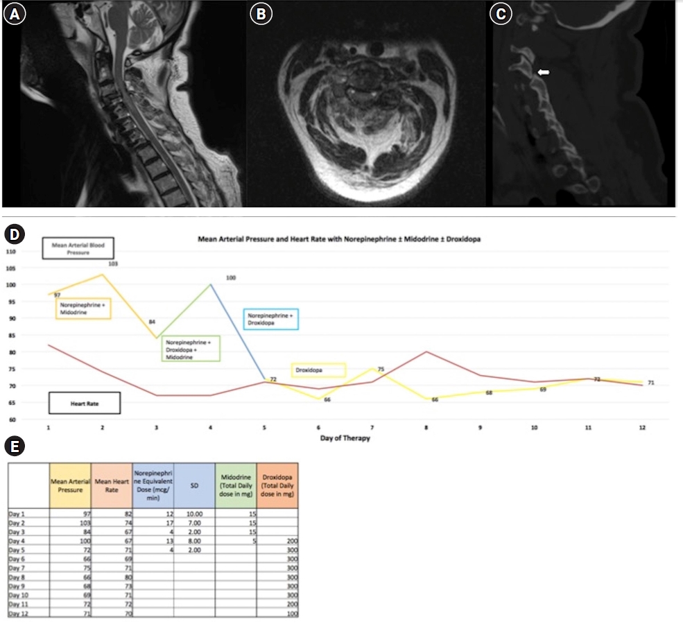
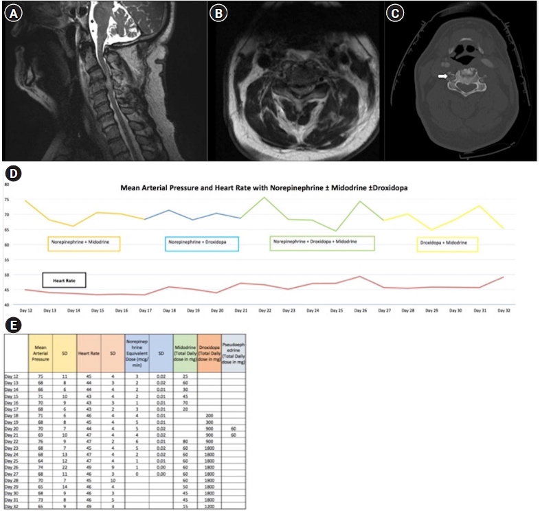
Acute traumatic spinal cord injury (SCI) is often associated with bradycardia and hypotension secondary to autonomic dysregulation. Secondary injury from hypotension is the most significant risk factor for poor outcomes in traumatic SCI [1]. While intravenous (IV) vasopressors are commonly utilized to reach blood pressure goals, they require central venous access; enteral drugs can potentially reduce the time required for central access and its associated complications. The most common enteral therapy utilized for hemodynamic augmentation is midodrine, a pure α-1-adrenergic agonist, but its use is often limited due to reflex bradycardia, and there are no established guidelines on its administration after SCI [2]. Droxidopa, an enteral precursor of norepinephrine, exerts its effects independently of the central nervous system and is used to treat neurogenic orthostatic hypotension (NOH) but has not been previously described to support blood pressure augmentation after acute SCI [3]. We present two cases of SCI with refractory hypotension for which we used droxidopa because of midodrine-induced bradycardia.
This was a single institution, retrospective review of patients who suffered acute SCI and were treated with droxidopa for hemodynamic augmentation. This study was carried out under a protocol, approved by the Institutional Review Board at Yale University. Informed consent from patients was waived.
Case 1
A 64-year-old male suffered a C3–4 fracture dislocation injury after falling 10 feet, resulting in paralysis of his lower extremities, right upper extremity, and minimal function of his left upper extremity (Figure 1A-C). He underwent urgent C3–4 laminectomies and posterior C3–6 fusion. Postoperatively, the patient failed to respond to aggressive fluid resuscitation to meet a target mean arterial pressure (MAP) goal >85 mm Hg, in accordance with guidelines after acute cervical SCI [4]. A tertiary trauma survey revealed no other significant injuries to aggravate hemodynamic instability from hypovolemia. As such, IV norepinephrine was started to maintain this MAP goal for 5–7 days and treat the neurogenic shock. On post-injury day 2, midodrine was started via a nasogastric tube at 10 mg three times daily (TID), to facilitate maintenance of the MAP goal while weaning norepinephrine. The patient continued midodrine for 4 days but could not be weaned off norepinephrine to maintain a MAP >85 mm Hg and experienced several episodes of significant bradycardia, resulting in midodrine discontinuation. Droxidopa was subsequently started on post-injury day 5 at a dose of 100 mg TID; in the setting of clinical improvement with improved activation of the proximal upper extremity muscles, the MAP goal was liberalized to >65 mm Hg. IV norepinephrine was successfully discontinued within 24 hours of droxidopa initiation. Droxidopa was continued for a total of 8 days, during which the MAP averaged 70 mm Hg without dropping below 65 mm Hg. Additionally, during this time the patient underwent tracheostomy and percutaneous endoscopic gastrostomy tube placement, secondary to his high cervical injury. The patient was transitioned to a lower level of care within 72 hours of droxidopa initiation. At the time of discharge, droxidopa was discontinued, and his neurological exam was unchanged from presentation. Details of IV and enteric vasopressor administration are detailed in Figure 1D and E.
Case 2
A 73-year old male with existing severe cervical spinal stenosis suffered a bicycle crash with hyperflexion injury causing paraplegia secondary to herniated C3–4 and C4–5 disc fragments (Figure 2A-C). He was able to extend his elbows against gravity, had trace movement in elbow flexion and finger grip, and could move both legs in the plane of the bed. Vital signs were notable for sinus bradycardia (40 bpm) and relative hypotension (MAP of 65 mm Hg) that did not improve with fluid resuscitation. After an urgent evaluation by cardiology, he was started on norepinephrine to facilitate blood pressure augmentation in the setting of neurogenic shock. He underwent urgent decompressive laminectomies at C3–5 with posterior fusion. Postoperatively, norepinephrine was continued for a goal MAP >85 mm Hg for 7 days. This was maintained successfully with norepinephrine, which was later transitioned to phenylephrine. He was also successfully extubated and was able to tolerate per os (PO) intake after a formal swallow evaluation. However, after 7 days, the phenylephrine could not be weaned off without MAP dropping below 65 mm Hg. On post-injury day 12, midodrine was started and up-titrated to total daily dose of 70 mg; this resulted in several bradycardic episodes. On post-injury day 18, droxidopa was started at 100 mg twice daily (BID) and up-titrated over several days to 300 mg TID. A pacemaker was considered but deferred in favor of monitoring his response to increasing doses of droxidopa. A brief trial of pseudoephedrine was performed without any clear benefit. Nearly 3 weeks after his initial injury, midodrine was restarted at 20 mg TID in addition to droxidopa 600 mg TID. He was weaned off norepinephrine by post-injury day 27. By post-injury day 32, midodrine and droxidopa were down-titrated to 5 mg TID and 400 mg TID, respectively. The patient was discharged to rehabilitation on this regimen with an improved neurological exam of antigravity strength in elbow flexion and extension bilaterally as well as antigravity strength in the right lower extremity. At last follow-up, 4 months after presentation, he remained on droxidopa therapy to maintain normotension (i.e. MAP >60 mm Hg. Details of IV and enteric vasopressor administration are detailed in Figure 2D and E.
Midodrine is often utilized to facilitate weaning IV vasopressors in the intensive care unit (ICU) setting. Midodrine acts only on the α1-receptor, causing increased peripheral vascular tone that may treat autonomic insufficiency and postural hypotension [5]. However, significant reflex bradycardia may occur in up to 15% of patients requiring vasopressor support in the ICU that may preclude its use [6,7]. Droxidopa is currently only approved by the U.S. Food and Drug Administration to treat NOH [3]. Droxidopa undergoes rapid bioactivation into norepinephrine, resulting in a mean MAP increase of approximately 40 mm Hg 1–3.5 hours after administration with effects lasting up to 8 hours [8]. Limited reports have described droxidopa to treat NOH after SCI. Wecht et al. [9] found increases in supine blood pressure with 200 and 400 mg doses of droxidopa in volunteers with NOH after chronic SCI. More recently, Canosa-Hermida et al. [10] described a patient with complete cervical SCI who developed NOH 6 weeks after the injury and whose symptoms abated with 300 mg BID of droxidopa.
In this report, we describe the novel use of droxidopa to treat refractory hypotension in the acute post-injury period in SCI patients who were unable to tolerate weaning of IV vasopressors. Notably, droxidopa’s conversion to norepinephrine occurs primarily in non-neuronal cells, and therefore its autonomic effects remains preserved in SCI [3,8]. Furthermore, baroreceptor mediated reflex bradycardia seen with pure α-agonists such as midodrine and phenylephrine has not been reported with the use of droxidopa, which would be expected based on its mechanism of action [3]. In both of our cases, midodrine alone could not successfully wean off IV vasopressors due to reflex bradycardia in the setting of increased vagal tone from known autonomic dysfunction. The addition of droxidopa, either as a monotherapy or combination therapy with midodrine, facilitated cessation of vasopressors (and avoidance of pacemaker placement in one patient). Notably, the daily dosages of droxidopa required for this wean varied greatly between the two patients, ranging from 300 mg to 1,800 mg—the latter being the maximum recommended daily dose [3]. However, even at high doses, the side effects reported for this drug in clinical trials were mild and included headache, dizziness, fatigue, and nausea [11]. Lastly, droxidopa may facilitate earlier weaning of IV vasopressors and subsequently earlier transfer out of the ICU, which may have significant resource and cost-saving potential for hospitals.
The indication for droxidopa in our patients was an inability to wean off vasopressors with midodrine. Currently, there are no well-accepted guidelines for use of midodrine, let alone droxidopa, for blood pressure augmentation after acute SCI. In our patients, the required dosing and timed response to droxidopa remained variable: patient 1 responded within 2 days to relatively low doses, whereas patient 2 required gradual up-titration of the drug over almost 2 weeks. Likewise, the required duration of droxidopa therapy remains unclear, as patient 2 still required droxidopa therapy at last follow-up, 4 months after presentation, to maintain normotension while patient 1 was weaned off droxidopa prior to hospital discharge. Current recommended droxidopa dosing is 100 mg TID, with up-titration every 24 to 48 hours to a maximum dose of 600 mg TID to treat NOH [3]. Higher doses may be needed in the setting of SCI but will require further investigation. Some of these questions may be answered in an ongoing early phase clinical trial, studying the dose response to droxidopa in hypotensive individuals with SCI (NCT03602014).
In summary, we report two cases of the use of droxidopa for hemodynamic augmentation after SCI. Droxidopa may be considered as an enteral agent to augment MAP in patients with autonomic dysfunction from acute TSCI and facilitate weaning of IV vasopressors. It may be a more optimal option than midodrine for SCI-related hemodynamic pathology due to less risk of bradycardia and hypothetically, may be particularly suited to patients with pre-existing cardiac co-morbidities. This strategy may avoid pacemaker placement and liberalize patients who are stable, but require continued admission in the intensive care unit for IV vasopressors, which can be cost ineffective and human resource depleting. Further cases studies are necessary to define the parameters and dosages for which droxidopa administration may be indicated in patients experiencing hemodynamic instability from autonomic dysfunction after SCI.

CONFLICT OF INTEREST

No potential conflict of interest relevant to this article was reported.

FUNDING

None.

AUTHOR CONTRIBUTIONS

Conceptualization: CSH, MKE, AAA, MLL. Data curation: all authors. Formal analysis: CSH, MKE, AAA, MLL. Methodology: CSH, AAA, MLL. Project administration: CSH, MKE, AAA, MLL. Visualization: CSH, MKE, AAA, MLL. Writing–original draft: all authors. Writing–review & editing: all authors.

None.
Figure 1.
Imaging, daily vital signs, and vasopressor doses of patient 1. Representative (A) sagittal and (B) axial images from T2-weighted magnetic resonance imaging of the cervical spine demonstrates severe C3–4 stenosis with associated cord signal. (C) Sagittal imaging from computed tomography shows jumped right-sided C3–4 facets, as indicated by an arrow. (D) Graphic and (E) tabular representations of daily mean arterial pressure, mean heart rate, and vasopressor dosing are shown. SD: standard deviation.
acc-2021-01662f1.jpg
Figure 2.
Imaging, daily vital signs, and vasopressor doses of patient 2. Representative (A) sagittal and (B) axial images from T2-weighted magnetic resonance imaging of the cervical spine demonstrates severe C3–4 and C4–5 stenosis with associated cord signal. (C) Axial imaging from computed tomography shows a comminuted fracture of the C3 transverse process, as indicated by an arrow. (D) Graphic and (E) tabular representations of daily mean arterial pressure, mean heart rate, and vasopressor dosing are shown. SD: standard deviation.
acc-2021-01662f2.jpg
  • 1. Ryken TC, Hurlbert RJ, Hadley MN, Aarabi B, Dhall SS, Gelb DE, et al. The acute cardiopulmonary management of patients with cervical spinal cord injuries. Neurosurgery 2013;72 Suppl 2:84-92.ArticlePubMedPDF
  • 2. Al-Abdouh A, Haddadin S, Matta A, Jabri A, Barbarawi M, Abusnina W, et al. Impact of adjuvant use of midodrine to intravenous vasopressors: a systematic review and meta-analysis. Crit Care Res Pract 2021;2021:5588483. ArticlePubMedPMC
  • 3. Keating GM. Droxidopa: a review of its use in symptomatic neurogenic orthostatic hypotension. Drugs 2015;75:197-206.ArticlePubMedPDF
  • 4. Walters BC, Hadley MN, Hurlbert RJ, Aarabi B, Dhall SS, Gelb DE, et al. Guidelines for the management of acute cervical spine and spinal cord injuries: 2013 update. Neurosurgery 2013;60:82-91.PubMed
  • 5. Brignole M, Moya A, de Lange FJ, Deharo JC, Elliott PM, Fanciulli A, et al. 2018 ESC guidelines for the diagnosis and management of syncope. Eur Heart J 2018;39:1883-948.ArticlePubMed
  • 6. Rizvi MS, Trivedi V, Nasim F, Lin E, Kashyap R, Andrijasevic N, et al. Trends in use of midodrine in the ICU: a single-center retrospective case series. Crit Care Med 2018;46:e628-33.ArticlePubMed
  • 7. Santer P, Anstey MH, Patrocínio MD, Wibrow B, Teja B, Shay D, et al. Effect of midodrine versus placebo on time to vasopressor discontinuation in patients with persistent hypotension in the intensive care unit (MIDAS): an international randomized clinical trial. Intensive Care Med 2020;46:1884-93.ArticlePubMedPMCPDF
  • 8. Kaufmann H, Saadia D, Voustianiouk A, Goldstein DS, Holmes C, Yahr MD, et al. Norepinephrine precursor therapy in neurogenic orthostatic hypotension. Circulation 2003;108:724-8.ArticlePubMed
  • 9. Wecht JM, Rosado-Rivera D, Weir JP, Ivan A, Yen C, Bauman WA. Hemodynamic effects of L-threo-3,4-dihydroxyphenylserine (Droxidopa) in hypotensive individuals with spinal cord injury. Arch Phys Med Rehabil 2013;94:2006-12.ArticlePubMed
  • 10. Canosa-Hermida E, Mondelo-García C, Ferreiro-Velasco ME, Salvador-de la Barrera S, Montoto-Marqués A, Rodríguez-Sotillo A, et al. Refractory orthostatic hypotension in a patient with a spinal cord injury: treatment with droxidopa. J Spinal Cord Med 2018;41:115-8.ArticlePubMed
  • 11. Kaufmann H, Freeman R, Biaggioni I, Low P, Pedder S, Hewitt LA, et al. Droxidopa for neurogenic orthostatic hypotension: a randomized, placebo-controlled, phase 3 trial. Neurology 2014;83:328-35.ArticlePubMedPMC

Figure & Data

References

    Citations

    Citations to this article as recorded by  

      • PubReader PubReader
      • ePub LinkePub Link
      • Cite
        CITE
        export Copy
        Close
        Download Citation
        Download a citation file in RIS format that can be imported by all major citation management software, including EndNote, ProCite, RefWorks, and Reference Manager.

        Format:
        • RIS — For EndNote, ProCite, RefWorks, and most other reference management software
        • BibTeX — For JabRef, BibDesk, and other BibTeX-specific software
        Include:
        • Citation for the content below
        Use of droxidopa for blood pressure augmentation after acute spinal cord injury: case reports
        Close
      • XML DownloadXML Download
      Figure
      We recommend

      ACC : Acute and Critical Care