A Rare Case of Massive Hemothorax due to Central Venous Catheterization Treated with Angiographic Stent Implantation
Article information

Abstract
In critically ill patients, centeral venous catheterization is a widely used procedure for fluid resuscitation, massive transfusion, total parenteral nutrition, central venous pressure monitoring and hemodialysis. However, many complications are associated with central venous catheterization. Among these complications, hemothorax is rare but fatal. We recently experienced a 32-year-old female diagnosed with hemothorax due to subclavian catheterization who was successfully treated with angiographic intervention. There are no absolute indications of surgery or interventional treatment in such cases. Multicenter studies and consensus are necessary to determine the proper treatment for hemothorax due to central venous catheterization. Angiographic treatment is rarely used for this uncommon complication of subclavian catheterization. We describe a rare case with a review of the literature.
Central venous catheterization is widely used procedure. In critically ill patients, centeral venous catheterization is needed for fluid resuscitation, massive transfusion, total parenteral nutrition, central venous pressure monitoring and hemodialysis. But, many complications are developed in central venous catheterization. These complications include pneumothorax, hemothorax, hematoma, hydrothorax, hydromediastinum, air embolism, thrombosis, infection and myocardial puncture.[1] Among these complications, hemothorax is rare but, fatal complication when developed. We recently experienced a 32-year-old female diagnosed with hemothorax due to subclavian catheterization and treated with angiographic intervention. This complication of subclavian catheterization is rare and angiographic treatment is not only difficult but also rarely used. We describe such a rare case and treatment with a review of the literature.
Case Report
A 32-year-old female presented to a local medical center with ongoing vaginal bleeding after normal vaginal delivery. The initial blood pressure was 90/60 mmHg and heart rate was 110/minute in the emergency room. Initial laboratory findings revealed Hb at 5.1 g/dL. Fluids and oxytocin were immediately supplied through peripheral intra-venous route. A subclavian catheterization was needed for vigorous fluid resuscitation, central venous pressure monitoring and transfusion. Right subclavian catheterization was first tried by emergency physician without sonographic guidance. During 18 gauge aspiration needle puncture, aspirated blood was pulsatile and fresh red. Therefore, puncture site compression was performed for several minutes. A second left subclavian catheterization was performed successfully. After cervix and perineal evaluation by obstetric physician, perineal obstetric wound was repaired. She received 7 units of red blood cells and 3 units of fresh frozen plasma. In follow up laboratory findings, Hb was increased to 12.8 g/dL. However, approximately 4 hours later, she developed confused mentality and dyspnea. Her blood pressure and heart rate were 80/40 mmHg and 120/minute respectively. In a third follow up laboratory findings, her Hb was decreased to 8.0 g/dL. Emergent orotracheal intubation was performed. Immediate chest radiograph was evaluated. Chest radiograph revealed haziness in right lung field (Fig. 1) suggesting possible hemothorax. Emergent tube thoracotomy was performed. Initially about 1000 cc blood was drained from chest tube. Dyspnea and haziness in radiograph were improved. Although fluid resuscitation was performed, blood pressure was 100/60 mmHg and pulse rate was 110/minute. After initial blood drainage from the chest tube, additional drained amount was about 1000 cc for 1 hour suggesting possible ongoing bleeding. Emergent angiography was performed by cardiovascular team. Angiography revealed subclavian artery bleeding into the thoracic cavity in angiography (Fig. 2). Therefore, stentgraft (Gore® VIABAHN® Endoprosthesis 8.0) was implanted. After implantation of stent graft, angiography showed no more bleeding (Fig. 3). After 4 days from angiography, extubation of orotracheal tube was performed. On hospital days 7, chest tube removal was performed. After that, the patient’s course was uneventful. She was discharged on hospital days 21.
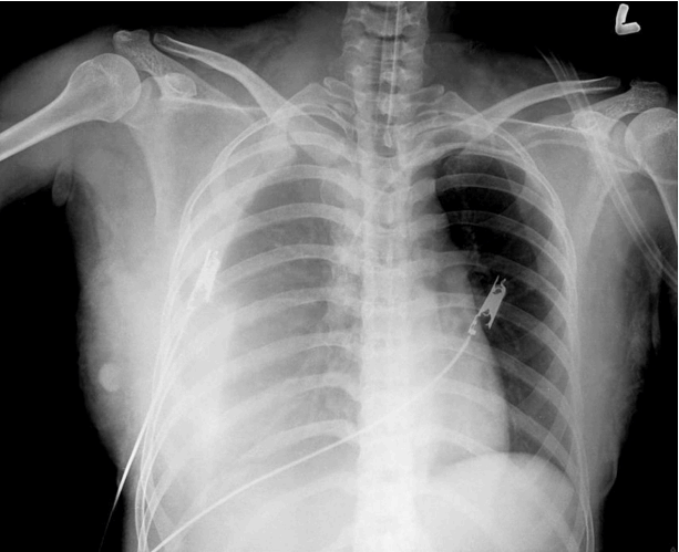
Chest radiograph. The chest radiograph shows a large hemothorax in the right thoracic cavity.
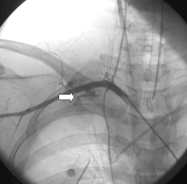
Angiography. Angiograpy shows extravation (white arrow) of contrast material into the thoracic cavity.
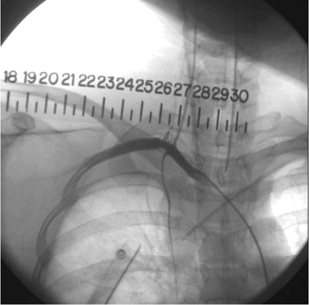
Angiography after implantation of a stent graft. There is no extravasation of constrast material after implantation of a stent graft.
Discussion
In critically ill patients, central venous catheterization was important and essential bedside procedures. Indications of central venous catheterization in critically ill patients include hyperalimentation, massive fluid resuscitation, transfusion, vaso-active drug use, central venous pressure or pulmonary wedge pressure monitoring and hemodilalysis.
In most cases, the central venous catheterization was performed safely without complication. However, in some cases, minor or lethal complications can develop. The complications of central venous catheterization include pneumothorax, hemothorax, hematoma, hydrothorax, hydromediastinum, air embolism, thrombosis, infection and myocardial puncture.[1] The total incidence rate of life-threatening complications during central venous catheterization is estimated at 0.02~1.5%.[2-4]
Hemothorax due to artery injury is one of the lethal complication. Artery injury rate in central venous catheterization is about 1-15%.[5,6] When artery injury occurs mortality can develop although adequate treatment was performed. [7,8]
In most cases, hemothorax is developed during insertion of catheter. However, during removal of catheter, hemothorax can also develop.[9] Therefore, care should be taken not only during insertion but also during removal of catheter.
Ultrasonography guided subclavian vein catheterization reduces the incidence of complications.[10] However, ultrasonography guided subclavian vein catheterization has not been widely used in clinical practice.[11] The causes of inactiveness in ultrasono-guidance subclavian vein catheterization include obesity, clavicle shape and anatomic position.[11]
In massive hemothorax due to subclavian artery injury, treatment options are surgery and interventional methods. In the anethesiology department, most hemothorax due to subclavian artery injury occur in the operating room with rescued patients immediately receiving surgical treatments.[12] In other cases, interventional methods instead of surgical treatment are used repair of artery injury depending on the lesion type. Interventional treatment methods include balloon tamponade, embolization, stent-graft placement and percutaneous closure devices.[13] In our case, stent implantation was performed successfully. The patient was completely cured without any complication. Because our cardio-vascular team was always stand-by state, prompt angiographic treatment was performed immediately. However, there are no absolute indications of surgery or interventional treatment. Although arterial puncture was developed, if small hematoma and hemodynamically stability were detected, close observation was necessary. A sudden deterioration of the general condition of the patient following a central venous catheterization should arouse suspicion of complications. In principle, prompt diagnostic procedures, such as chest radiograph, chest ultrasonography, trans-esophageal echocardiography, or chest computed tomography are needed. However, in the presence of a typical clinical scenario, it would be justified to perform immediate treatment without any time wasted in imaging evaluation.[7] In massive hemothorax due to subclavian atery injury, prompt resuscitation and adequate treatment are urgently needed without time consumed hesitation.
In conclusion, central venous catheterization is one of the frequently performed bedside procedures in critically ill patients. We describe a rare case with a review of the literature so that lethal complication of central venous catheterization such as hemothorax and its adequate treatment could be understood. Multicenter study and consensus are necessary to decide the proper treatment for hemothorax due to central venous catheterization.
Notes
No potential conflict of interest relevant to this article was reported.