Effective Postoperative Use of Dexmedetomidine in a Child with Severe Pulmonary Arterial Hypertension Secondary to Congenital Ventricular Septal Defect
Article information
Abstract
Although α2-adrenoceptor agonists are widely used as postoperative sedatives in adults, the postoperative effects in pediatric patients with secondary pulmonary arterial hypertension (PAH) due to congenital heart disease are not well known. We experienced a case of successful ventilator weaning with continuous intravenous administration of dexmedetomidine (DEX) after surgical correction in a 46-month-old child with congenital ventricular septal defect (VSD) with severe PAH. She underwent VSD closure on cardiopulmonary bypass (CPB). After successful weaning from the CPB, hemodynamics and oxygenation were stabilized on DEX and nitroglycerin in the intensive care unit. The patient was successfully weaned from the ventilator 46 hours after surgery. The transthoracic echocardiogram two weeks after surgery showed a closed VSD with no residual shunt and trivial tricuspid regurgitation (Vmax = 2.5 m/sec) without PAH.
Fatal postoperative pulmonary arterial hypertension (PAH) crisis can be caused by excessive stimulation of the sympathetic nervous system, and this is one of the cardiovascular complications that is observed in patients with PAH secondary to congenital heart disease (CHD) after corrective heart surgery. PAH has been shown to be a significant predictor of major perioperative cardiovascular complications in patients undergoing cardiac catheterization or non-cardiac surgery under anesthesia, including PAH crisis, cardiac arrest, and death.[1] The use of α2-adrenoceptor agonists as a postoperative sedative has been well-established in adult patients. However, the effect of dexmedetomidine (DEX) administration after heart surgery in pediatric patients with secondary PAH due to CHD has not been well studied to date. Here we describe a case of successful ventilator weaning with continuous intravenous administration of DEX after the surgical correction of a ventricular septal defect (VSD) in a child with severe PAH secondary to congenital VSD.
Case Report
A 46 month-old female with body weight of 8.56 kg and height of 84 cm (body surface area: 0.45 m2) was admitted for evaluation and treatment due to orthopnea, exertional dyspnea, growth retardation, and poor feeding since infancy. The left chest had a bulged appearance due to cardiomegaly. Tremor of the entire chest was felt on palpation. On auscultation, low grade pan-systolic murmur was heard across the entire left chest. Chest X-ray showed increased pulmonary vascularity, signs of pulmonary edema, and cardiomegaly (Fig. 1). A preoperative transthoracic echocardiogram (TTE) revealed a large perimembranous VSD with a diameter of 1.5 cm and a left-to-right (LR) shunt of 1-1.5 m/sec velocity, severe PAH with dilated main pulmonary artery (PA), and mild TR of 4-4.5 m/sec velocity (Fig. 2). Medical therapy was initiated with diuretics, digitalis, and a Ventavis® inhalator (Actelion Pharmaceuticals, Switzerland). After one week of medical therapy, a follow-up TTE was performed. LR shunt velocity had increased to 2.3-2.5 m/sec, and tricuspid regurgitation velocity was diminished to 3.7-3.8 m/sec. Surgical closure of the VSD was performed through a median sternotomy using a Sauvage patch using cardiopulmonary bypass (CPB) with myocardial protection by intermittent hypothermic antegrade crystalloid cardioplegia and maintenance of mild systemic hypothermia. After successful weaning from the CPB under the support of a Ventavis® inhalator, the patient was transported to the intensive care unit (ICU) in stable condition. Upon ICU admission, Midazolam® (Bukwang Pharmaceuticals, Korea), Perdipine® (Astellas Pharmaceuticals, Japan), and Tridol® (Yuhan Co., Korea) were administered once. Intravenous dexmedetomidine (DEX, 0.2-0.5 μg/kg/h) and nitroglycerin (NTG, 0.25-0.5 μg/kg/min) were continuously administered with intermittent intravenous analgesia (Tridol®). Upon sedation, hemodynamics and arterial blood gasses were stabilized, and the patient was smoothly and successfully weaned from ventilator support within 46 hours after surgery. After weaning, the doses of both drugs were gradually reduced and discontinued before ICU discharge. Two weeks after surgery, TTE showed that the VSD was closed without residual shunt, and showed trivial TR (Vmax = 2.5 m/sec) without significant PAH (Fig. 3). A chest X-ray performed 2 weeks postoperatively (Fig. 4) showed diminished heart size and no signs of pulmonary edema. The patient was discharged from the hospital in good condition.
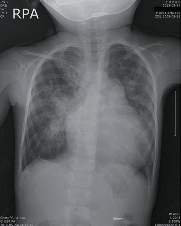
Chest X-ray on admission. Increased pulmonary vascularity, signs of pulmonary edema, and cardiomegaly. RPA: right posterior anterior view.
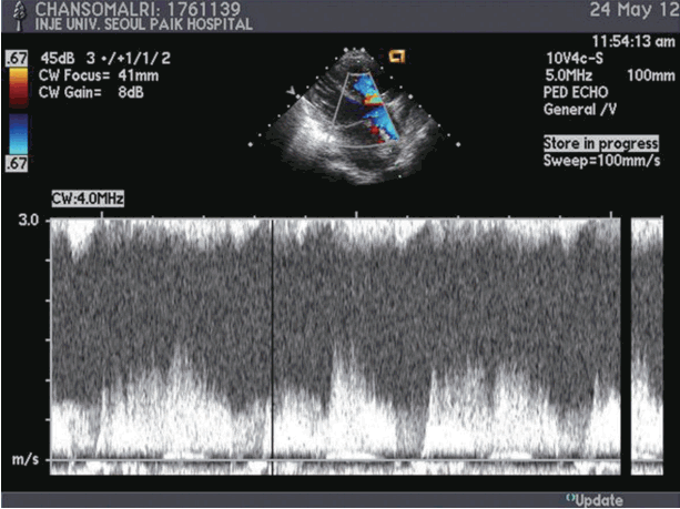
Preoperative TTE. Large perimembranous VSD with a diameter of 1.5 cm and a left to right shunt (Vmax = 1-1.5 m/ sec). TTE: transthoracic echocardiogram; VSD: ventricular septal defect; CW: continuous wave doppler; PED ECHO: pediatric echocardiography.

Postoperative TTE, two weeks after surgery. Closed VSD with no residual shunt, and trivial TR (Vmax=2.5 m/sec) with no significant PAH. TTE: transthoracic echocardiogram; VSD: ventricular septal defect; TR: tricuspid regurgitation; PAH: pulmonary arterial hypertension; CW: continuous wave doppler; PED ECHO: pediatric echocardiography.
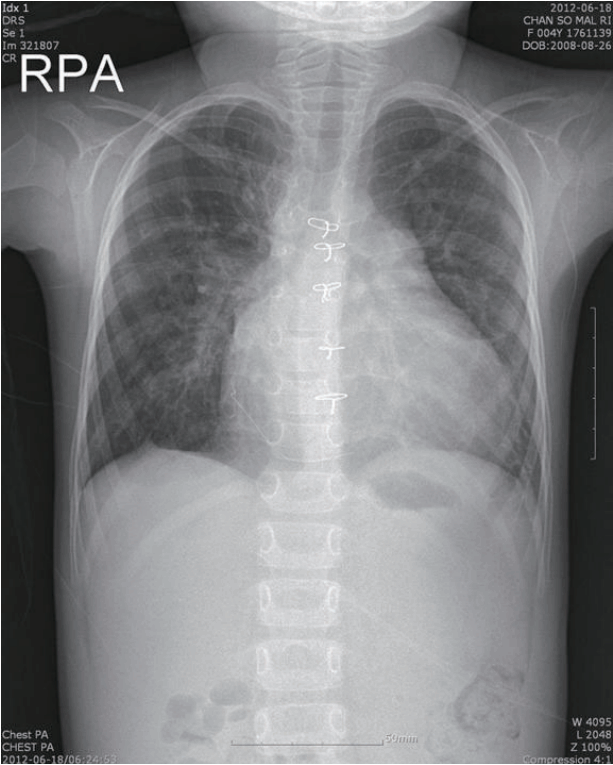
Postoperative chest X-ray, two weeks after surgery. No signs of pulmonary edema, and diminished heart size. RPA: right posterior anterior view.
Discussion
A sudden increase in pulmonary vascular resistance can cause an increase in right ventricle (RV) afterload, RV dysfunction, and hemodynamic decompensation, resulting in PAH crisis. In turn, PAH results in inadequate pulmonary blood flow, inadequate left ventricular preload, low cardiac output, and biventricular failure. The consequent hypotension causes coronary ischemia, which becomes a vicious cycle. Hypoxia, hypercarbia, acidosis, hypothermia, pain, and airway manipulations are considered to be factors affecting development of PAH crisis.[1] Respiratory depression due to opioid drugs can also affect PA pressure in spontaneously breathing patients.
DEX is likely to have beneficial effects in PAH patients. It acts as a specific central α2-adrenoceptor agonist in the locus ceruleus, improving hemodynamics, and providing sedation by reduction of sympathetic tone. The ‘arousable sedation’ induced by DEX allows the patient to respond to verbal commands. Its moderate analgesic effects are mediated via α2 receptors in the dorsal horn cells of the spinal cord; it acts synergistically with opioids. Slow intravenous loading is recommended, as rapid loading can cause systemic hypertension via direct action on peripheral α2, ß receptors in the arteriolar resistance vessels.[2] At low-dose infusions of DEX, respiratory depression is minimal[3] and cardiopulmonary function in healthy volunteers is well maintained at doses that provide adequate sedation.[4] It considerably reduces the incidence of emergence agitation in children who are undergoing sevoflurane anesthesia, and bolus administration at 1 μg/kg has been observed to be safe, without increased side effects.[5]
Administered intraoperatively, DEX causes attenuation of the hemodynamic and neuroendocrinal response to surgical trauma and CPB in pediatric patients who are undergoing corrective surgery for congenital heart disease.[6] DEX-based combination therapy not only provides optimal sedation, but it also prevents and ameliorates fatal morbidities after pediatric cardiovascular surgery, as is also seen to be the case in adults.[7] However, little is known regarding the postoperative use of this drug in pediatric patients with PAH secondary to CHD, although use has been demonstrated in the pediatric ICU as an effective sedative in children with chronic neurologic impairments.[8] The one retrospective study evaluating the prolonged use (more than 24 hours) of DEX in the pediatric cardiothoracic ICU did not observe significant adverse effects with respect to any of the clinical outcome examined, and found that it reduced the need for other sedatives and narcotics.[9]
The recommended standard dose of a 0.5 μg/kg bolus over 10 min followed by an infusion of 0.5 μg/kg/h would decrease in the lower dose ranges as described[4] and it might not have a direct vasoconstrictive effect. However, a study of the extended use of DEX in adults also found that administering the drug as an infusion, and avoiding boluses, abolished problematic hemodynamic effects.[10] DEX-induced arousable sedation and moderate analgesia resulted in improved oxygenation with hemodynamics. In pediatric patients with PAH who are at high risk for postoperative cardiovascular complications following cardiovascular surgical procedures, DEX may enable sufficient sedation in conjunction with hemodynamic stabilization and minimal effect on the pulmonary circulation.
Notes
No potential conflict of interest relevant to this article was reported.