Clozapine Induced Neuroleptic Malignant Syndrome
Article information

Abstract
Neuroleptic malignant syndrome is a rare, but potentially life-threatening adverse event associated with the use of neuroleptic agents. We describe the case of a 47-year-old schizophrenic woman who was treated with clozapine for years. The patient developed acute renal failure with pulmonary edema, and underwent mechanical ventilation and hemodialysis.
Neuroleptic malignant syndrome (NMS) is an uncommon, but potentially life-threatening side effect of antipsycotic medications.[1] NMS is a known idiosyncratic and unpredictable adverse reaction that is related to the administration of dopamine antagonists.[2] We report a case of NMS in a schizophrenic patient who was treated with clozapine for years prior to the onset of NMS.
Case Report
A 47-year-old Korean woman was diagnosed with schizophrenia and treated with clozapine for years, from 2004 onward. It was determined through outpatient follow-up appointments that the patient had poor adherence to her prescribed medication regimen. Additionally, the patient and her spouse had divorced within the past year. The patients’ mother passed away 5 months preceding the event. The patient did not have a regular caregiver; however, 3 days prior to the adverse event she moved in with her son. While she was able to communicate with her son until the day preceding the event, she was drowsy and incoherent when found at home.
When she arrived at the emergency department, azotemia (blood urea nitrogen, 29 mg/dL; creatinine, 2.56 mg/dL), muscle enzyme elevation (creatine kinase, 70,758 IU/L; lactate dehydrogenase, 933 IU/L), and gross hematuria on urinalysis with metabolic acidosis (pH, 7.28; PaCO2, 33 mmHg; HCO3- 15.5 mmol/L) were identified through laboratory tests. Anuria was verified over several hours. Under a clinical diagnoses of rhabdomyolysis, the treatment plan included hemodialysis and mechanical ventilation to address the patient’s respiratory distress that had developed as a result of pulmonary edema (Fig. 1). At that time, leukocytosis (white blood cell count, 31,940 cells/μL), high acute phase reactant (C-peptide protein, 7.04 mg/dL), and sustained fever (38.0oC) were observed, all of which suggested the development of an infection. In consideration of sepsis, systemic antibiotics were administered. After admission to the intensive care unit (ICU), continuous renal replacement therapy was administered because of low blood pressure requiring inotropic agents, and was continued for days. As a result, the pulmonary edema improved. Nevertheless, extubation was successful on the fourth day of ICU admission (Fig. 2), drowsiness persisted, and a generalized tonic-clonic seizure occurred. No abnormalities were found on the brain magnetic resonance image and then the electroencephalogram showed no epileptiform discharge. Furthermore, we performed cerebrospinal fluid analysis but there was no evidence of infection in the cerebrospinal fluid, except for the presence of elevated protein levels (61 mg/dL). Organisms were not isolated from the cerebrospinal fluid. An antiepileptic drug was prescribed, and the seizure-like movement subsided.

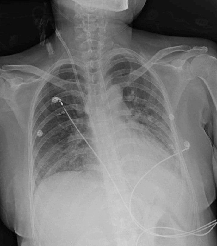
A chest radiograph reveals prominent bilateral vascular markings suggestive of acute pulmonary edema.
A chest radiograph reveals improved bilateral pulmonary edema compared with a chest radiograph taken 3 days prior.
Appropriate antibiotics were administered for several days following the urine culture, proving that extendedspectrum beta-lactamase-resistant Escherichia coli induced urosepsis. Leukocytosis did not improve despite the decreased C-reactive protein levels and although the fever subsided. She was prescribed clozapine again after regaining consciousness. After resuming clozapine treatment, the leukocytosis became severe, from 24,970 cells/μL to 72,070 cells/μL over the course of 6 days and fever was reappeared (37.8oC) but there was no definite evidence of systemic infection on blood test and culture study and fever was responsive to antipyretic drug.
The possibility of primary hematologic disease is highly unlikely because of the absence of atypical leukocytes on the peripheral blood smear examination, and a normal range of white blood cells on a blood test that was reported a month before. In accordance with severe systemic inflammation and under the suspicion of underlying myeloproliferative disease, short-term systemic steroids were administered for 5 days, without clinical improvement and the result of serum BCR/ABL1 rearrangement test was negative.
While there was no bottle of medicine around her at the time that she was found, her will was uncovered within the days following the adverse event. Under the circumstances, it is likely that the patient attempted suicide by overdosing on clozapine and clinically NMS caused by clozapine overdose was suspected. Under the suspicion of NMS induced by clozapine, we discontinued clozapine administration, after which, the leukocytosis rapidly resolved to the normal range, from 72,070 cells/μL on the day of the last dose of clozapine was administered to 7,000 cells/μL 7 days after clozapine was withheld.
Because the patient was on a clozapine regimen for several years, an idiosyncratic reaction is less likely. It is strongly suggestive that an overdose could have led to clozapine-induced NMS.
After the continuous renal replacement therapy was switched to intermittent hemodialysis, the patient was transferred from the ICU to the general ward, and a low-dose of clozapine was resumed to control her delusional symptoms.
Discussion
NMS is a potentially life-threatening condition if not correctly diagnosed. This condition is associated with the use of neuroleptic agents. Recent studies suggest that patient mortality is estimated to range between 10% and 20%.[3]
Other drugs that induce dopamine receptor blockade for agitation, sedation, or emesis could cause the development of NMS. Regarding the type of antipsychotic drug, typical antipsychotics administered at a higher potency are associated with a higher risk for the development of NMS. Nonetheless, newer, less potent atypical antipsychotics including clozapine, olanzapine, risperidone, and quetiapine could cause NMS.[4-6]
NMS has been reported to occur at all doses and routes of administration.[4-6] Several studies have suggested that NMS is likely to occur more frequently during the initial months of treatment, after a rapid increase in dosage, or following parenteral injection.[7,8] Studies report that NMS may occur at any time during the course of medication administration, even after a patient has been treated with the same agent, at the same dosage.[2,9] In our patient, she has taken clozapine for years but she takes overdose with suicidal intent, overdose could able to cause NMS.
The clinical features of NMS include fever, rigidity, mental-status changes, and autonomic instability.[2] The fever is usually very high, without fluctuations, and does not respond well to antipyretics.[4] Rigidity is usually generalized and symmetric, but focal increase in muscular tone may also be present.[4] Mental status changes include delirium, changes in the level of consciousness, disorientation, and agitation. Autonomic instability presents as tachycardia and labile blood pressure.[2] Creatine kinase elevation >10,000 IU/L is typical, and a relatively specific laboratory indicator used in the diagnosis of NMS. Increased levels of creatine kinase reflect more advanced stages of the disease and a poor prognosis.[10,11] Other laboratory abnormalities that are commonly seen are not as specific, and include leukocytosis; elevated levels of lactate dehydrogenase, liver transaminase, and inflammatory markers such as C-reactive protein; and increased erythrocyte sedimentation rate.[2,12] In the present case, CK elevated upto 63,046 IU/L which implicate advanced stage and poor prognosis but finally her CK decreased to 39 IU/L and discharged alive without any persistent organ failure. Rhabdomyolysis resulting in acute renal failure with myoglobinuria may also be observed as in this case.[5]
A single presenting sign must be identified in over 80% of cases, and NMS may occasionally have a fulminant onset.[13] Furthermore, there are no confirmed diagnostic criteria for NMS yet. A collection of detailed information about drug history coupled with laboratory abnormalities helps the clinician to arrive at a diagnosis.[2] A broad range of disorders that manifest as fever should be considered in the differential diagnosis of NMS, and medical and neurologic evaluations are needed. To exclude primary disorders of the brain that may resemble NMS, such as structural lesions and infectious encephalitis, brain imaging studies and lumbar puncture are required.[ 4,12] An electroencephalography may be needed to exclude epilepsy, in rare cases.[4] Routine muscular biopsy is not recommended for diagnosing NMS.[2]
Withholding the causative agent is the most important factor in the treatment of NMS. Other treatment options mainly comprise supportive care.[2] Therefore, a clinician should be vigilant and attempt to diagnose NMS as early as possible to consider the appropriate rationale for the interruption or continuation of neuroleptics.
In conclusion, we report a case of NMS in a patient with schizophrenia who had been on a clozapine regimen for years, without dose escalation. In this case, without a high suspicion of NMS, the patient was subject to misdiagnosis. Because of the unpredictability of NMS, when we deal with a patient who is taking neuroleptics and showing signs of NMS, we need to rule out conditions that may show similar symptoms. Clinicians should consider disorders that present similar symptoms and distinguish NMS, and thus offer appropriate care and prevent potentially fatal complications.
Notes
No potential conflict of interest relevant to this article was reported.